Abstract
Pregnancy causes many changes in a woman’s body. The growing foetus causes mechanical changes by stretching skin, muscle, and fascia. The amount of physiological change during pregnancy varies among women but often persist long after the post-partum period despite the best efforts of the patient regarding exercise and diet.
Many women find it difficult to deal with these major body changes and this article theorises why humanoid females are almost unique amongst species in accumulating fat in the bitrocanterea (saddlebag or culotte) area and lower abdomen midriff, while males seem more prone to develop visceral fat around their organs? It also notes how during pregnancy human skin often stretches out of shape and loses the ability to spring back into shape forming sagging breasts and stretch marks in the period. Lastly it looks at other changes such as pigmentation and varicose veins and what patients should do to get the best aesthetic result and get their bodies back to a pre-pregnancy state. Many of the examples and treatments mentioned are based on the personal preference and experience of the author.
Pregnancy leads to many changes in a woman’s body, mainly through the interaction of steroid hormones, lactogen, and cortisol on the underlying tissues and structures1. The growing foetus itself causes mechanical change by stretching skin, muscle, and fascia and demanding an increased calorific supply2. The amount of extra weight gained during pregnancy varies among women3. The National Health Service recommends that the overall weight gain during the 9 month period for women who start pregnancy with normal weight should be in the region 10 to 12 kg (22–26 lbs) but as many British females are already over weight these figures may be downsized. Physicians are aware, insufficient weight gain can compromise the health of the foetus during pregnancy and excessive weight gain can pose risks to the woman and the baby. For women who have gained weight between pregnancies, even a relatively small gain of 1–2 body mass index (BMI) units can increase the risk of high blood pressure or diabetes during their next pregnancy and may also increase the chance of giving birth to a large baby. New advice has just been published by the National Institute for Health and Clinical Excellence (NICE) as part of its public health programme4.
It states that women who are obese (with a BMI over 30) when they become pregnant face an increased risk of complications such as diabetes, miscarriage, pre-eclampsia, blood clots, and death. Obese women are also more likely to have an induced or longer labour, post-delivery bleeding, and slower wound healing after delivery. They also tend to be less mobile, which can result in a need for more pain-relieving drugs during labour. These can be difficult to administer in obese women, resulting in a greater need for general anaesthesia with its associated risks4.
So why do women have such a hard time dealing with this major body change, which often persists long after the post-partum period despite their best efforts regarding exercise and diet? Why do females develop layers of subcutaneous fat around their midriff, their buttocks, and inner thighs while males seem more prone to develop visceral fat around their organs? Why do pelvic bone widening changes occur in humans often causing major distress resulting in long term urinary and bladder dysfunction? Why does the skin stretch out of shape and lose the ability to spring back into shape?
Adipose tissue
For many years, there was little interest in the biochemistry or physiology of adipose tissue. It is now well recognized that adipocytes play an important dynamic role in metabolic regulation. They are able to sense metabolic states via their ability to perceive a large number of nervous and hormonal signals. More recent insights show that adipocyte secretion itself can have a major metabolic impact5. The breakdown of cellular fat stores fuels energy production and multiple anabolic processes. Albert et al, demonstrated that the lack of hormone-sensitive lipase, a member of the enzyme trio that catabolizes fat, has pronounced effects on lipid metabolism, glucose homeostasis, and cell signaling in humans6. Associations have been made by Karine Clement and others between genetic obesity susceptibility and early postnatal fat and lean mass7.
Bariatric surgery represents a powerful tool for morbid obesity treatment. It is usually used for patients with a BMI of around 40 or more (extremely obese). However, after the stabilization of weight loss that follows surgical interventions, ex-obese patients face the problem of residual tissues removal. It is unknown whether the characteristics of this residual subcutaneous adipose tissue (SAT) is ‘restored’ with regard to molecular and morphological features8.
One of the more interesting theories to explain why female fat deposits are located where they are was made by a Polish researcher called Boguslaw Pawlowski of the University of Wroclaw. To quote from an issue of Current Anthropology ‘the fat deposits may help to meet the balance requirements of two-legged walking during pregnancy and lactation’9.
In this publication, Pawlowski notes that both during advanced pregnancy and when nursing, a human female has an additional anterior load that moves her centre of gravity forward and upward, making bipedal locomotion more difficult and energetically inefficient. This theorises why we see a lot of the problems we see in the post-natal period. We have a quadruped means of reproduction been carried around in a biped body. Therefore, evolution may have promoted buttocks and thigh fat deposits to compensate for the biomechanical handicap imposed by carrying a baby. Interesting theory but it would probably explain some of the cultural effects of this new shape. We hence have two physiological types:
The ‘pre-baby’ or ‘mating’ body type with fat distribution seen in more ‘normal’ areas of distribution. In this body type, breasts are again used to ‘attract’ a partner rather than being a functional mammary organ. They tend to be ‘firmer’ due to fat distribution. Women prefer this body type as it symbolises health, vitality, and beauty. Men prefer it as it shows a female who is ready to reproduce.
The ‘post-baby’ body type with fat now distributed subcutaneously especially in the buttocks and thigh area. There are breast volume changes with sagging, especially if the female has breast fed her infant. Breast volume may increase but usually we get shrinkage. In this physiological state the dermal tissue of the abdomen has been distended and sometimes left with long-term ‘stretch marks’.
We shall now look at the problem encountered by the ‘post-baby’ body type and what a patient can do to correct them. In some instances the tissues have been distended and sheared to such an extent that surgical repair is the best if not the only option. We will first look at excess fat. The author does not put dieting and exercise as a primary option because most patients feel it is not effective.
Excess fat
Solution: Liposuction (tumescent, ultrasonic-assisted, power-assisted, water-assisted, and laser-assisted) bariatric surgery
Liposuction, also known as lipoplasty (‘fat modeling’), liposculpture suction lipectomy (‘suction-assisted fat removal’) or simply lipo, is a cosmetic surgery operation that removes fat from many different sites on the human body. Areas affected can range from the abdomen, thighs, and buttocks, to the neck, backs of the arms and elsewhere10. There are many differing mechanism of liposuction and the procedure has been reported to be associated with significant morbidity and risk of mortality under general anaesthesia11. Conventional liposuction consists of using large cannulae under general anaesthesia, largely practiced by plastic surgeons. The whole procedure lasts 2–3 hours. This method is quick, can remove large amounts of fat, and saves time for the surgeon. It is performed under general anaesthesia and only a small amount of fluid is introduced into the fat. It is possible to remove large amounts of fat, often 8–10 litres (called megaliposuctions), quickly in 1–2 hours, through large cannulae (6 mm-1 cm in diameter).
However, it has many disadvantages, including the fact that the patient has to be hospitalized, which adds significantly to the cost and the possibility of hospital-acquired infections. It goes without saying that general anaesthesia always has its risks. There is a risk of side‑effects, such as fat embolism, which can be potentially fatal. Large cannulae need large incisions which have to be sutured and heal with significant scars12. The use of large cannulae also causes greater damage to tissue and, hence, increases the bleeding. This technique is associated with significant blood loss, often needing blood transfusions13. Recovery time is slow, as after any procedure under general anaesthesia.
Microcannular tumescent liposuction involves subcutaneous infiltration of large volumes of crystalloid fluid called Klein’s solution, which contains low concentrations of lignocaine and epinephrine, followed by suction-assisted aspiration of fat by using small aspiration cannulae called microcannuale. The term tumescent liposuction specifically excludes the use of any additional anaesthesia, either intravenous or gaseous, and by definition, is a method for performing liposuction surgery with the patient totally under local anaesthesia14,15. Since the first description by Jeffrey Klein, dermatologic surgeons have made significant contributions in this field, and tumescent liposuction using microcannuale under local anaesthesia, is regarded as safe and effective. There have been many developments in this field16, including those mentioned below:
- Suction-assisted liposuction (SAL)
- Ultrasound-assisted liposuction (UAL)
- Power-assisted liposuction (PAL)
- Twin-cannula (assisted) liposuction (TCAL or TCL)
- External ultrasound-assisted liposuction (XUAL or EUAL)
- Water-assisted liposuction (WAL)
- Laser Assisted.
Liposuction can be combined with other procedures (i.e. radiofrequency) that involve a level of skin retraction. The level of skin retraction following liposuction is affected by the age of the patient, quality of skin, presence of underlying disease or smoking, and the presence of previous skin damage such as those caused by childbirth and surgery. Surgical lifts are also used post-pregnancy to address massive weight loss when the combination of large amounts of skin and shrunken fat cause significant skin drooping. Large volume Liposuction (SAL) in combination with other surgery is common, but may have higher complication rates.
Non-liposuction alternatives
The risks, financial costs, and lengthy downtime associated with surgical procedures for fat reduction have led to the development of a number of non-invasive techniques. Non-invasive body contouring now represents the fastest growing area of aesthetic medicine17. There are currently four leading non-invasive techniques for reducing localized subcutaneous adipose tissue: low-level laser therapy (LLLT), cryolipolysis, radiofrequency (RF), and high-intensity focused ultrasound (HIFU).
Cryolipolysis is the non-invasive cooling of adipose tissue to induce lipolysis — the breaking down of fat cells — to reduce body fat without damage to other tissues. It initially received FDA clearance for fat reduction in the abdomen and flanks, and more recently there has been significant interest in non-surgical fat reduction for other sites, such as the inner and outer thighs. Zelickson and Burns reported results of an inner thigh study that contributed to FDA clearance of cryolipolysis for the treatment of thighs18.
The author believes, without question, the development of SAL/ultrasound-assisted liposuction (UAL) changed the face of excisional body contouring surgery as many plastic surgeons use it an adjunct to excisional abdominoplasty. Most physicians use this system to remove fat from common areas such as the abdomen, waist, hips, back, buttocks and thighs, as well as more delicate areas such as the arms, calves, ankles, knees, face and neck19. Some physicians also use the device to sculpt female breasts. A majority of doctors questioned in a recent survey found that the system offered the following benefits over traditional liposuction19:
- Faster patient recovery
- Less pain medication required
- Minimal bruising
- Reduced need for re-treatments
- Skin tightening
- Increased precision.
Preliminary findings from a multi-centre clinical study measuring skin retraction in ultrasound-assisted liposuction patients shows 40% to 60% skin tightening. Most doctors agree that the key to good skin retraction is related to treating the layer of fat directly under the skin. This allows the physician to effectively sculpt an area and stimulate the dermal collagen, resulting in skin tightening19. Another study in the July/August issue of the Aesthetic Surgery Journal, found that this device significantly limited blood loss during liposuction when compared to traditional liposuction alone. The authors concluded that ultrasound-assisted lipoplasty should be recommended over traditional suction-assisted lipoplasty for patients undergoing large-volume liposuction procedures or treatments in very fibrous areas of the body where increased blood loss is expected20.
Another recent development is the use of Nutational Infrasonic Liposculpture (NIL) or ‘tickle’ liposculpture. The procedure was originally developed in Europe and was approved by the FDA for use in the US in 2010. It uses low frequency acoustic infrasonic vibration combined with simultaneous suction to loosen and remove unwanted areas of fat from the body. Unlike traditional liposuction, NIL uses air pressure rather than heat, which causes less damage to surrounding tissue and blood vessels. The cannula used to remove the fat is also much narrower, resulting in less bruising and scarring after the procedure. NIL can be used on any area of the body previously treated with traditional liposuction methods. The procedure is quickly gaining popularity as doctors and patients alike are comparing the benefits it offers over traditional lipoplasty procedures.
Healthy eating habits combined with regular exercise also help people lose weight. However, this natural process, possibly as a result of Pawlowski’s theories above, takes more time and determination after pregnancy. Weight loss via exercise and healthy eating carries little risk compared to liposuction.
Stretch marks
Solution: Carboxytherapy, radiofrequency, photodynamic therapy, dermarolling, TCA peeling
Stretch marks occur during pregnancy and are caused by rapid stretching of the dermal tissue of the abdomen during the distension and weight gain of pregnancy21. It is thought that nearly 85% of women will develop some degree of stretch marks during their pregnancy and these usually appear after prolonged weakening of the dermal tissues about the beginning of the third trimester. This is also a period of sustained distension22.
It is felt that dermal weakening occurs during pregnancy as a result of increased glucocortoid hormones, which actually effect the epidermis formation of fibroblasts23. The reduced numbers of fibroblasts consequently result in less collagen and elastin being formed and this leaves the dermal tissue supporting structure susceptible to tearing. These skin changes eventually result in reddish or purple lines along the planes of stretching that tend to gradually fade to a lighter skin colour as a tendency towards re-epithelialisation occurs. Over time they can diminish but they do not disappear completely and can cause distress in the post natal female. There is little doubt that stretch marks are also influenced by hormonal changes in pregnancy as is also seen during the rapid growth of puberty or weight gain during muscle building24.
Various treatments are available for the purpose of improving the appearance of existing stretch marks, including laser treatments, dermabrasion, and prescription retinoids. In terms of lasers the author has used 585 nm pulsed dye lasers with some effect but prefers a combination of dermarolling with radiofrequency. There are many publications citing this means of therapy25, and the author finds bipolar and tripolar devices to be of the most benefit. The use of RF brings about many questions, the most important is which type of collagen is induced, and if the result of the action is a fibrotic tissue. If fibrotic tissue is being formed to increase the firmness of the skin, then it won’t help to recover the physiology of the connective tissue and the result will be short-lived and not beneficial in the long term.
More recently, The author has treated abdominal stretch marks with fractionalised CO2 laser resurfacing with some effect. This method is limited by little published data but the author has treated approximately 15 cases with lower density settings over multiple treatments in order to wound the dermis and try and recreate the collagen and elastin underlying structure, which the skin was deprived off. One can expect about 70% benefit over five sessions spaced 1 month apart26.
Many patients ask if there are any topical creams to help with stretch marks, either during their formation or after they have occurred. There are no proper control studies but some research suggests a daily application of a cream containing Gotu Kola extract, vitamin E, and collagen hydrolysates was associated with fewer stretch marks during pregnancy27. Other favourites include combinations of cocoa butter, vitamin E, panthenol, hyaluronic acid, elastin, and menthol. It was associated with fewer stretch marks during pregnancy versus no treatment27. In some cases we have almost complete resolution with Carboxytherapy and this is the author’s preferred treatment.
Carboxytherapy is a simple and proven technique that can dramatically improve the appearance of stretch marks by improving local tissue metabolism and perfusion. Treatments are rapid, comfortable, and effective for a high percentage of patients. Carbon dioxide (CO2) is infiltrated into the subcutaneous tissue through a tiny 30 G needle (0.3 mm in diameter)28. From the injection point, the CO2 diffuses easily into adjacent tissues. Unfortunately, nothing much has been published in the literature about this technique, except for its relationship to break down adipose tissue29. Various treatments have been proposed for the treatment of post-pregnancy stretch marks but in the author’s opinion most of them are not as good as carboxytherapy.
Sagging skin
Solution: abdominoplasty
Abdominoplasty or ‘tummy tuck’ is a popular cosmetic surgery procedure used to make the abdomen firmer after pregnancy. The surgery (which can be radical) involves the removal of excess skin and fat from the middle and lower abdomen in order to tighten the muscle and fascia of the abdominal wall. Abdominoplasty operations vary in scope and are frequently subdivided into categories, depending on the extent of the surgery. There are different variations on the technique dependent on which tissues are sagging and if there is associated weight loss or residual fat to be removed. The first variant is whether a partial or a complete abdominoplasty is required30.
- Complete abdominoplasty can take from 1 to 5 hours. During this procedure, an incision is made from hip to hip just above the pubic area and another is made to free the navel from the surrounding skin. The skin is then detached from the abdominal wall to reveal the muscles and fascia to be tightened
- Partial abdominoplasty (mini-tuck abdominoplasty) can be completed between 1 to 2 hours. During this technique, a smaller incision is made and the skin and fat of the lower abdomen are detached in a more limited fashion from the muscle fascia. The skin is stretched down and excess skin removed
- Extended abdominoplasty includes a lateral thigh lift. The operation allows for complete abdominal contouring as well as smoothing the contour of the upper lateral thigh
- High lateral tension abdominoplasty (HTLA) tightens abdominal muscles in a vertical line. This method, in addition to vertical-line tightening, tightens muscles horizontally. The final result with this technique is a dramatically flat abdomen with significantly better-defined waistline31
- Floating abdominoplasty or FAB technique (also known as an extended mini-abdominoplasty) allows for tightening and shaping through a smaller incision that isn’t placed around the belly button. Through this smaller incision, excess skin is removed and the belly button is temporarily detached, floating above the muscles during this process. The muscles are tightened and reshaped from sternum to pubic area.
- Combination abdominoplasty includes a lower body lift done in conjunction with another procedure such as breast reduction, breast lift, hysterectomy or liposuction contouring.
Abdominoplasty carries certain risks that may be serious or life-threatening. When making the decision to undergo such a procedure it is recommended to compare the benefits with the potential risks and complications. Hence, all patients must be informed of all the risks they are exposing themselves to32. These combination techniques mean the patient is under anaesthesia for a longer period of time, increasing the mortality risk. Contraindications to abdominoplasty include right, left, or bilateral upper quadrant scars (relative); severe comorbid conditions (eg, heart disease, diabetes, morbid obesity, cigarette smoking); future plans for pregnancy (relative); a history of thromboembolic disease (relative); morbid obesity (BMI >40); and unrealistic patient expectations. Combined aesthetic and gynecologic surgery is an attractive option for both patients and surgeons. A popular name for breast enhancement procedures performed in conjunction with an abdominoplasty after pregnancy is now called a ‘Mommy Makeover’.
Breast atrophy
Solution: Breast implants and hyaluronic acid
This is one of the most obvious places to see the difference between a ‘pre’ and ‘post’ pregnancy body type. During pregnancy, the ovaries and the placenta produce estrogen and progesterone. These hormones stimulate the 15 to 20 lobes of the milk-secreting glands in the breasts to develop. It is widely accepted that most breasts seem to undergo a ‘deflationary’ change in the weeks and months after weaning. The reason that this occurs is due to the fact the ratio of fat tissue to gland tissue in a women’s breast actually changes with the new requirements of the body. Most women tend to regain their normal cup size post natally but many do not, especially if they have breast fed. The body has underwent a physiological redistribution of fat and when the breast gland tissue regresses there is less fat and the breast may shrink, especially in the upper poles. Ptosis of the breast is the medical term for drooping or sagging female breasts. Many women and medical professionals mistakenly believe that the breast itself offers insufficient support and that wearing a bra prevents sagging. Many also believe that nursing increases sagging. Society is hard on mothers and very few magazine pictures display this new physiotype as anything to be proud of.
One of the most popular treatments for breast atrophy remains breast implants. There are three general types of these devices, defined by their filler material: saline solution, silicone gel, and composite filler. The saline implant has an elastomer silicone shell filled with sterile saline solution; the silicone implant has an elastomer silicone shell filled with viscous silicone gel; and the alternative composition implants featured miscellaneous fillers, such as hyaluronic acid, soy oil and even polypropylene string33. When compared to the results achieved with a silicone-gel breast implant, the saline implant can yield acceptable results of increased breast-size, smoother hemisphere-contour, and realistic texture; yet, it is likelier to cause cosmetic problems, such as the rippling and the wrinkling of the breast-envelope skin, accelerated lower breast pole stretch, and technical problems, such as the presence of the implant being noticeable to the eye and to the touch34.
Women with breast implants may have functional breast-feeding difficulties; mammoplasty procedures that feature periareolar incisions are especially likely to cause breast-feeding difficulties. Surgery may also damage the lactiferous ducts and the nerves in the nipple-areola area.
The presence of radiologically opaque breast implants (either saline or silicone) might interfere with the radiographic sensitivity of the mammograph, that is, the image might not show any tumour(s) present. In which case, an Eklund view mammogram is required to ascertain either the presence or the absence of a cancerous tumour, wherein the breast implant is manually displaced against the chest wall and the breast is pulled forward, so that the mammograph can visualize a greater volume of the internal tissues. Nonetheless, approximately one-third of the breast tissue remains inadequately visualized, resulting in an increased incidence of mammograms with false-negative results35.
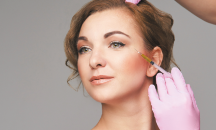
Hyaluronic acid injection treatment has now largely been discontinued in the UK and Ireland as a breast enhancer due to the lack of consensus amongst radiologists regarding how to examine breasts that have been injected with filler. The technique involved injecting stabilised hyaluronic acid into the deflated breast and then moulding to the desired shape. The benefits are that it requires only a local anaesthetic and will leave no scarring, although bruising, swelling and discomfort for a few days are expected. The effect however only lasts for 12 months, after which further injections are required. The procedure has also drawn criticism as its long term effects are relatively unknown. The only medical trial was supported by the manufacturers, involving 1000 patients in Japan, and anybody taking the procedure will be entered into a European-wide research trial. Problems with this treatment included capsular formation and product migration. The author witnessed some patients with small lumps of product that required aspiration.
The breast augmentation patient is usually a young woman whose personality profile indicates psychological distress about her personal appearance and her bodily self-image, and a history of having endured criticism about the aesthetics of her person. In 2008, a longitudinal study conducted by Lipworth et al, reported that women who sought breast implants are almost 3.0 times as likely to commit suicide as are women who have not sought breast implants36.
Breast sagging
Solution: Breast lift
Breast lift (mastopexy) refers to cosmetic surgery designed to lift or change the shape of a person’s breasts. The surgery may involve repositioning the areola and nipple, as well as lifting the breast tissue and removing skin37. A breast lift may be performed alone, or in combination with placement of breast implants; when implants are used, the procedure is typically called breast augmentation. The effects of a breast lift are often reduced with time as the shape and distribution of existing breast tissue tends to be temporary, as the effects of gravity and ageing continue, causing ptosis to recur over time38. A classification system has been suggested by Regnault and modified by numerous authors39. The most commonly used system is as follows:
- Grade 1: Mild ptosis — nipple just below inframammary fold but still above lower pole of breast
- Grade 2: Moderate ptosis — nipple further below inframammary fold but still with some lower pole tissue below nipple
- Grade 3: Severe ptosis — nipple well below inframammary fold and no lower pole tissue below nipple; ‘Snoopy nose’ appearance.
Pseudoptosis is inferior pole ptosis with nipple at or above inframammary fold; usually observed in postpartum breast atrophy.
Women who experience multiple pregnancies repeatedly stretch the skin envelope during engorgement while lactating. As a woman’s breasts change in size during repeated pregnancies, the size of her breasts change as her mammary glands are engorged with milk and as she gains and loses weight with each pregnancy. In addition, when milk production stops (usually as a child is weaned), the voluminous mammary glands diminish in volume, but they still add bulk and firmness to the breast. These changes in the mammary glands contribute further to sagging.
Melasma
Solution: Azelaic acid (20%), hydroquinone, tretinoin, intense pulsed light, fractional CO2 laser
Melasma is the formation of irregular pigmented patches and are commonly found on the sun exposed face in the period surrounding pregnancy. It is thought they are caused by increasing levels of both estrogen and progesterone and may be seen outside pregnancy during administration of the oral contraceptive pill40. The hormones are thought to stimulate melanocytes resulting in increased production of the normal tanning protective chemical but why this pigment occurs in patches or even gets caught in the dermis is less well known. There appears to be a racial predisposition to the problem, especially in East Europeans and Jews41. It is also known to occur in patients with thyroid disease and stress may result in increased levels of melanocyte stimulating hormone42. The pigmentation usually disappears itself four months after the circulating hormone levels return to normal.
The author feels it is important that someone with clinical experience of pigmented lesions should be involved in their removal. There are many reasons for this, melanomas can regress and depigment and lesions such as lentigo malignas can appears in the same locations on the face as melasma. That said, melasma is comparatively easy to diagnose as it tends to occur in large patches rather than isolated lesions. When there is doubt, the author recommends using a Wood’s UV light to determine the depth of melanin pigmentation in the skin: contrast in epidermal pigmentation is increased while contrast in dermal pigmentation is decreased under Wood’s lamp illumination compared to ambient visible light. Under Wood’s lamp, excess melanin in the epidermis can be distinguished from that of the dermis.
Dermal melanin is more difficult to remove by methods that do not reach this level. Of particular benefit is a dermal melanin analysis system but not all clinics have this technology at their disposal. The author finds this is of particular benefit, especially when using a series of IPL laser treatments to help depigmentation. If the pigmentation is dermal then another method of removal will be required as it will only serve to darken the lesions. Whenever dermal melanin is involved the author will use a fractionalised CO2 laser. Mandelic acid has also been shown to be of benefit in these cases.
If the melasma is epidermal then topical depigmenting agents, such as hydroquinone (HQ) are a good starting point. These creams come in either in OTC (over-the-counter) (2%) or POM prescription (4%) strength. As physicians, we tend to use 4% as standard. Hydroquinone is a chemical, which inhibits tyrosinase, one of the enzymes involved in the production of melanin.
The author also uses azelaic acid (20%) as it also decreases the activity of melanocytes. Hydroquinone topical has been assigned to pregnancy category C by the FDA. Animal studies have not been reported. There are no controlled data in human pregnancy. Hydroquinone topical is only recommended for use during pregnancy when benefit outweighs risk. It is unknown if hydroquinone cream is excreted in breast milk.
After pregnancy, the author uses many topical creams for depigmentation. Tretinoin as 0.025% OTC or 0.05% POM is a retinoic acid that increases skin cell (keratinocyte) turnover. This treatment cannot be used during pregnancy. After pregnancy, facial peels with alpha or beta hydroxy acids or chemical peels with glycolic acid can also be used. The biggest problem with removal of melasma is the possibility that the condition will return. The patient should also be told the lightening effects are gradual because the pigmented cells have to grow out to the stratum corneum and a strict avoidance of sunlight post procedure is mandatory. We should also be aware that ordinary sunscreen in my opinion does not prevent melasma recurring and topical agents with physical blockers, such as titanium dioxide or zinc dioxide should be used. Ordinary light will make melasma recur. Some products such as those containing vitamin C and SPF benefits provide some protection and nourishment to skin. Other products containing retinol, (a less irritating form of vitamin A), can boost cell turnover within the skin’s surface to reduce the appearance of dark spots. Other less wounding treatments such as glucosamine, mulberry root, and yeast extracts can work to accelerate the skin’s natural exfoliation process, in effect buffing away the dark spots. Of course the patient may not want a procedure and may be told that cosmetic cover-ups can also be used to reduce the appearance of melasma.
Varicose veins and spider veins
Solution: Lasers, IPL, sclerotherapy, surgery
Pregnancy tends to worsen spider veins and smaller varicose veins because circulating hormones associated with pregnancy soften the vein walls and valves. During pregnancy, veins have to carry a greater circulating blood volume. Sometimes the enlarged uterus compresses abdominal veins, causing further back pressure on the leg veins. Changes in body chemistry due to birth control pills and constrictive clothing, such as tight hosiery, can also contribute to spider vein development. Other related factors are heredity, obesity, menopause, ageing, prolonged standing, leg injury, and abdominal straining43. Treatment can be either conservative or active. Active medical intervention can be divided into surgical and non-surgical treatments. Newer methods including endovenous laser treatment, radiofrequency ablation, and foam sclerotherapy appear to work as well as surgery for varices of the greater saphenous vein44.
The medical term for spider veins is telangectasias. These fine red and purple veins are approximately 1 millimeter or less in size and can occur anywhere on the body. Patients most commonly seek treatment for spider veins on the legs and chest. Spider veins are often found in combination with varicose veins. Laser and IPL treatment of spider veins is accomplished using the 595 nm pulsed-dye laser, the 1064 YAG laser or IPL. These systems produce vein specific wavelengths which seal the spider veins45,46.
Sclerotherapy has been used in the treatment of varicose veins for over 150 years. The procedure is used to treat smaller varicose veins with a chemical sclerosing agent or type of foam in order to make these vessels necrose or shrink in size. In this rather simple procedure, veins collapse and fade from view. The procedure may also remedy the bothersome symptoms associated with spider veins, including aching, burning, swelling and night cramps. The medicines that are commonly used as sclerosants are polidocanol (POL), sodium tetradecyl sulphate (STS), glycerin and chromated glycerin and hypertonic saline47. The liquids can be mixed at varying concentrations of sclerosants and varying sclerosants/gas proportions, with air, CO2 or O2 to create foams.
A number of techniques have been performed for over a century, from the more invasive saphenous stripping, to less invasive procedures like ambulatory phlebectomy and CHIVA. Stripping consists of removal of all or part the saphenous vein (great/long or lesser/short) main trunk. The complications include deep vein thrombosis, pulmonary embolism and wound complications including infection48. There is evidence for the great saphenous vein growing back again after stripping.