


Oksana Pashkovska, MD, explores the latest techniques in under-eye injectables, addressing key challenges like volume loss, oedema, and the Tyndall effect to enhance patient safety and aesthetic outcomes
The periorbital region plays a crucial role in facial aesthetics and expression1. This area is central to interpersonal communication, as the eyes are often referred to as the ‘window to the soul’2. Changes in this region can significantly impact the overall appearance and aesthetic perception of the face.
With age, the periorbital area undergoes a series of anatomical and clinical transformations that profoundly affect facial aesthetics. One of the primary changes is subcutaneous fat atrophy, leading to volume loss and the formation of hollows under the eyes. This phenomenon results from the reduction and downward displacement of fat compartments due to gravitational forces3.
Furthermore, weakening of the ligamentous apparatus and periorbital muscles contributes to wrinkle formation, skin sagging, and the appearance of under-eye bags. Specifically, stretching and thinning of the orbital septum allows fat pads to protrude, creating visible swelling and puffiness4.
Another important factor is skin thinning in this area. As the skin loses elasticity and becomes more transparent, underlying structures such as blood vessels and muscles become more visible, contributing to the appearance of dark circles5.
Additionally, changes in the facial bony structures, particularly orbital rim resorption, lead to an increase in the orbital aperture and deeper-set eyes. This exacerbates the aesthetic deterioration of the periorbital area as soft tissues lose support and descend6.
The combination of these changes is influenced by both intrinsic ageing factors and external factors, such as ultraviolet radiation, smoking, and environmental pollution, which accelerate structural degradation and impact the overall aesthetic quality of the periorbital region.


Excess volume and the risk of ptosis
Filler injections into the periorbital area can lead to excessive volume, which, in turn, contributes to the development of ptosis. This phenomenon, known as facial overfilled syndrome, is characterised by the distortion of natural facial contours due to excessive or improperly placed fillers7.
Over time, under the influence of gravity and age-related changes, these excess volumes may further exacerbate tissue ptosis, particularly in the delicate periorbital region8.
A study by Mobin Master demonstrated that hyaluronic acid fillers can persist in tissues much longer than previously expected. Magnetic resonance imaging (MRI) revealed that fillers may remain at the injection site for several years, increasing the risk of volume accumulation and ptosis development9.
Tyndall effect
The Tyndall effect occurs when light scatters on filler particles that have been injected too superficially, resulting in a bluish or cyanotic discolouration of the skin. This phenomenon is particularly noticeable in the periorbital region due to the thinness of the skin10.
Incorrect injection technique or the use of an inappropriate filler can contribute to the development of this undesirable effect, which may persist for an extended period, making correction challenging11.
Oedema
Hyaluronic acid fillers have the ability to attract water, which can lead to oedema at the injection site. In the periorbital region, where the tissues are particularly sensitive, this can become a significant issue12.
Incorrect filler selection, excessive injection volume, or inappropriate injection technique can increase the risk of oedema. Additionally, the depth and area of filler placement play a crucial role in minimising this complication13.

Techniques
With age, a decrease in the maxillary angle is likely the cause for the expansion of the inferior orbital rim and, thus, of an anterior positioning of the orbital septum. Therefore, a pseudoprolapse of the intraorbital quarter retroseptal fat pads, fat compartments located behind the orbital septum in the lower eyelid, can lead to the palpebral bags becoming more apparent. Also, the orbicularis retaining ligament might lose its horizontal position toward a more inferior inclined alignment, causing a loss of stability to the adjacent orbicularis oculi muscle, which forms the anterior wall of the underlying fat compartments below the aperture, that is, the suborbicularis oculi fat (SOOF)14.
The injection technique described in the works of Casabona and Bernardini considers the ligamentous structure of the face to optimise lifting and volume restoration. This method involves injecting filler into a specific anatomical point, known as the G-point, located in the lateral portion of the SOOF. Filler placement in this area enhances midface elevation and improves the contour of the periorbital region. Studies have demonstrated that this technique achieves significant aesthetic improvement with minimal filler volume15.
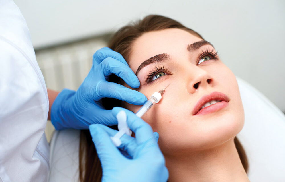
Therefore, as the first step in infraorbital correction for most patients, I perform filler injection into the dermal location termed the G-point16 (Figure 1). This point is determined by the intersection of three reference lines:
- A line connecting the inferior aspect of the nasal ala and the tragus
- A line connecting the lateral canthus and the corner of the mouth
- A connecting line between the intersection of the two lines and a perpendicular line connecting to the lateral canthus.
If there are indications for midface volume restoration, filler can also be injected using a cannula under the SMAS layer in a fan-shaped, linear retrograde technique along the zygomatic arch and into the projection of deep fat compartments. Alternatively, a bolus injection with a needle can be performed directly into the deep fat compartments and along the zygomatic bone (Figure 1B).
For the first step in infraorbital correction, I use Alexa Volume filler (Hyalual, Switzerland). The second stage of infraorbital correction involves working directly with the lower eyelid to reduce the transition between the eyelid and the cheek. While a cannula is generally considered safer, its use in the periorbital area may cause trauma to the orbital septum, damage to retaining ligaments, and injury to lymphatic capillaries, which can lead to complications.
Considering these factors, in some cases, the use of a needle for superficial subdermal filler injection along the orbital ligament is a more predictable and controlled approach (Figure 2).
The disadvantages of this technique include the potential for filler contouring and the Tyndall effect. Therefore, it is crucial to prioritise a filler that distributes evenly, is easy to mold, and has a low tendency for swelling.
In my practice, for this area, I use Alexa Medium filler (Hyalual, Switzerland), which meets these requirements. Due to its phosphate-buffered saline composition and rheological properties, Alexa has a volumisation factor of 1:1 (Figure 3).
The periorbital region is critically important for the aesthetic perception of the face, and its correction requires a deep understanding of the anatomical and physiological characteristics of this area. Further research and the development of new treatment methods will contribute to improving the effectiveness and safety of procedures aimed at enhancing the appearance of this delicate region.
Declaration of interest The author, Oksana Pashkovska, is a Key Opinion Leader (KOL) for Hyalual, but the views expressed are solely her own. Every effort has been made to provide an objective analysis of the product.


References
- Schlessinger J, Cohen JL. Periorbital rejuvenation: strategies for treatment. Dermatol Surg. 2015;41 Suppl 10:S143–S147.
- Carruthers J, Carruthers A. Aesthetic indications for botulinum toxin injections. Plast Reconstr Surg. 2003;112(5):23S–27S.
- Rohrich RJ, Pessa JE. The fat compartments of the face: anatomy and clinical implications for cosmetic surgery. Plast Reconstr Surg. 2007;119(7):2219–2227.
- Hwang K, Kim DJ, Hwang SH. Surgical anatomy of the orbital septum and periorbital fat pads. J Craniofac Surg. 2007;18(2):387–391.
- Wulc AE, Sharma P, Czyz CN. The anatomic basis of midfacial aging. Facial Plast Surg Clin North Am. 2015;23(2):191–202.
- Pessa JE, Chen Y. Curve analysis of the aging orbital aperture. Plast Reconstr Surg. 2002;109(2):751–755.
- Lim T. Facial overfilled syndrome. Dermatol Clin. 2024;42(1):121–128.
- Cotofana S, Lachman N. The facial overfilled syndrome: anatomy, causes, and prevention. Aesthet Surg J. 2022;42(4):343–350.
- Master M, Mobin MB. Hyaluronic acid filler longevity and localisation: magnetic resonance imaging evidence. Plast Reconstr Surg. 2021;147(1):50e–53e.
- Sundaram H, Fagien S. Considerations for hyaluronic acid fillers in the periorbital area. Plast Reconstr Surg. 2021;147(5):888–902.
- Lowe NJ, Lask GP, Yamauchi PS. Blue discoloration after hyaluronic acid filler injection: Tyndall effect and its management. J Cosmet Laser Ther. 2007;9(3):182–187.
- Swift A, Remington K. Periorbital edema and hyaluronic acid fillers: causes, prevention, and treatment. J Clin Aesthet Dermatol. 2020;13(8):55–62.
- Gold MH, Alam M, Burgess C. Avoiding and managing adverse effects of hyaluronic acid fillers in the periorbital area. Dermatol Surg. 2019;45(12):1582–1592.
- Cotofana S, Fratila AM, Schenck TL, Redka-Swoboda W, Zilinsky I, Pavicic T. The anatomy of the aging face: a review. Facial Plast Surg. 2016;32:253–260.
- Casabona G, Bernardini F, et al. How to best utilise the line of ligaments and the surface volume coefficient in facial soft tissue filler injections. J Cosmet Dermatol. 2019;18(6):1452–1460. DOI: 10.1111/jocd.13245.
- Casabona G, Bernardini FP, Skippen B, et al. How to best utilise the line of ligaments and the surface volume coefficient in facial soft tissue filler injections. J Cosmet Dermatol. 2019;00:1–9. DOI: 10.1111/jocd.13245.


