Erythema and elastometry
Skin erythema was measured using a II ColorMeter (Cortex Technology, Denmark). Elastometry values were measured using a Dermacheck Multi Skin Center MC750 (Courage-Khazaka Electronic GmbH Koln, Germany). All measurements were taken on standardised facial anatomical landmarks identified by the intersection between a vertical line passing though the centre of the pupil and a horizontal line touching the base of the columella on both cheeks. Erythema was measured first in order to avoid possible secondary vaso-reactivity induced by the suction probe used to measure elastometry. All measurements were taken at constant thermal comfort room parameters, immediately before the first treatment session (T1), and subsequently at 45 days (T2 - T1 + 45 days) and 75 days (T3 - T1 + 75 days).
Subject’s self-assessment
Simple questionnaires to evaluate subjective intra-operative perceived sensations were given to each subject at the end of the first treatment (T1–IOP) and at the end of the full eight treatment sessions (T1 + 30 days–IOP). Seven parameters were evaluated during LED exposure:

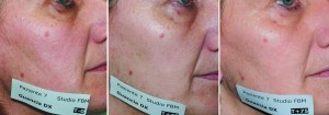
Figure 2 Clinical pictures taken at T0 (immediately pre-treatment), T45 (15 days after eight LED sessions), and T75 (45 days after eight LED sessions)
-
Pleasant feeling (PF)
-
Absence of pain (AOP)
-
Safety perception (SP)
-
Absence of fear (AOF)
-
Burning perception (BP)
-
Warmth perception (WP)
-
Claustrophobia perception (CP).
Four different sensations were evaluated immediately after the end of the first treatment (T1-END) and at the end of the complete treatment sessions (T1 + 30 days–END): pleasant feeling (PF); absence of pain (AOP); burning perception (BP); and warmth perception (WP). Five more subjective perceptions were evaluated 24 hours after the first treatment (T1-END + 24 hours) and after the complete treatment sessions (T1 + 30 days + 24 hours):
-
Not interfering with social activities (NISA)
-
Not interfering with make-up use (NIMU)
-
Absence of pain (AOP)
-
Absence of desquamation (AOD)
-
Absence of redness (AOR).
Each parameter was evaluated according to a five-point scale as follows: 1 = totally disagree, 2 = disagree, 3 = indifferent, 4 = partially agree, 5 = completely agree.
Subjective assessment of clinical results were obtained 15 days (T2 - T1 + 45 days) and 45 days after completion of full treatment sessions (T3 - T1 + 75 days). Study subjects were previously instructed on how to evaluate their skin tone improvement, wrinkle reduction, colour uniformity improvement, and skin texture improvement; thus, all parameters were subjectively evaluated on a five-point assessment scale as follows: 1 = totally ineffective, 2 = moderately effective, 3 = effective, 4 = very effective, and 5 = extremely effective.
Overall clinical outcome assessment
Clinical photography assessment was performed blindly by two independent dermatologists who were asked to identify the temporal order of digital pictures taken at T1: immediately pre-treatment; T2: T1 + 45 days (15 days after the completion of eight LED treatments); and T3: T1 + 75 days (45 days after the completion of eight LED treatments), after being blindly reshuffled by a nurse. A simple percentage match was used to evaluate the magnitude of agreement between the two reviewers.
Results
All ten subjects completed the full series of treatment sessions and attended all scheduled follow-up visits. No variations were detected regarding subjects’ BMI during the duration of the study.
Overall clinical outcome photographic assessment by independent reviewers
Both reviewers correctly identified the pre-treatment photographs (100% concordance). Moderate disagreement was registered with regard to the correct assignment of T2 (T1 + 45 days) and T3 (T1 + 75 days) images (72% concordance).
Objective assessment of skin texture, wrinkles, and superficial pigment alterations
Skin texture irregularities, wrinkle count, and superficial pigment irregularities were automatically evaluated using the computerised feature count system installed on a Visia Complexion Analysis unit. Computerised assessments were made immediately before the first LED treatment (T1), at T2 (T1 + 45 days), and at T3 (T1 + 75 days).
Global skin texture feature count was found significantly decreased from baseline values at T2 (-8.96%) (Figure 4) followed by a slight upward variation, keeping values still below baseline at T3 (- 6.93) (Figure 5). Global wrinkle feature count showed an even more marked decrease from baseline values at T2 (-23.23%), followed by a slight upward variation at T3 (-16.8). Superficial spot feature count showed an opposite global trend scoring a +0.69% at T2 and a + 11.96% at T3.
Erythema and elastometry
Surface skin erythema showed a slight increment at T2 (+ 0.97%) followed by a more pronounced increase at T3 (+ 4.81%), confirming the positive metabolic stimulation of LED treated regions (Figure 8). Global elastometry showed a marked improvement at T2 (+ 20.59%) followed by a moderate decrease (+ 10.99%), keeping still above baseline values, at T3.


