


Jana Janovska and Lana Kasparane discuss how obesity and metabolic syndrome can play a role in undermining skin barrier function, which can lead to a variety of adverse skin conditions

Department of Internal diseases, Riga Stradins University, Dermatovenerologist at Dermaclinic Riga and Capital Clinic Riga, Riga, Latvia; Lana Kasparane, Third year resident in Dermatovenerology, University of Latvia, Dermatovenerologist at Dermaclinic Riga, Riga, Latvia
Metabolic syndrome (MetS) is a condition in which the patient has a central form of obesity (women > 80 cm, men > 94 cm) and at least two of the following criteria — elevated triglyceride or glucose levels in the blood, low high-density lipoprotein (HDL) levels, and high blood pressure(1). Its prevalence in industrialised and developing countries is rapidly increasing(2).
In several international studies, various clinical skin manifestations that are associated with MetS and/or its components have been identified. The diseases include psoriasis, androgenetic alopecia, acanthosis nigricans, skin tags, lichen planus, acne inversa, systemic lupus erythematosus, and even skin cancer(3). But is it associated with sensitive skin? As we know, metabolic syndrome maintains an elevation of inflammatory cytokines and supports a chronic inflammatory reaction in the skin, so this relationship is possible. There is little information in the scientific literature about sensitive skin association with metabolic syndrome. However, knowing the fact that metabolic syndrome patients have constantly increased inflammatory parameters and, in the case of sensitive skin, immune cells are also activated, there could be connections.
Sensitive skin
Sensitive skin is a clinical condition that is observed in all economically developed and industrialised countries, with almost the same spread in America and Eurasia. Similar to MetS, it is still rapidly rising, which is a severe problem because of the proven adverse effects on the patient’s quality of life(4,5).
Sensitive skin manifests with the varying intensity of subjective indications, such as burning and soreness, as a response to factors like wind, heat, dryness, air conditioning, psychoemotional stress, hormonal disturbances, too frequent and/or improper skincare.
The pathophysiological mechanism is still not completely understood. Still, there are several theories that speculate on the cause of skin hypersensitivity, such as skin barrier changes, keratinocyte activation, neurogenic inflammation/neural mechanism, decreased skin tolerance and genetic predisposition. In the scientific field, the most often described and investigated possible pathogenesis mechanisms are skin barrier function changes and neurogenic inflammation. This is understandable because all these factors are interconnected and mutually complementary — impaired barrier function of the stratum corneum leads to the exposure of immune system cells and sensitive nerves, resulting in marked cutaneous responses to otherwise harmless stimuli(4–7).
“Skin is the largest organ in the human body. One of its main tasks is to protect against external harmful environmental factors.”
Skin barrier function and inflammation
Skin is the largest organ in the human body. One of its main tasks is to protect against external harmful environmental factors. The skin barrier can be divided into three levels of protection:
- physical barrier against pathogens, mechanical trauma and uncontrolled loss of water and dissolved substances (stratum corneum, epidermis cells with nuclei, and desmosomes)
- chemical/biochemical barrier with antimicrobial activity — innate immunity (lipids, organic acids, lysosomes, and antimicrobial peptides)
- humoral and cellular immune systems (lymphocytes, neutrophils, monocytes, and Langerhans cells).
There are multiple factors that can affect the integrity of the skin barrier, such as the humidity of the environment, pollution, UV radiation, increased age, stress, hypertension, and exposure to excessive water. It has also been found that morbidly obese patients have impaired skin barrier repair(8).
The skin barrier consists of keratinocyte differentiation and constant regeneration. During differentiation, keratinocytes undergo various stages of development. It is a very complex process involving proteins, fatty acids, lipids and enzymes. Their functions and expression are controlled by cytokines and intercellular signal molecules and receptors. So, to ensure the proper development of the epidermal barrier, the correct regulation of all components, which are involved in the differentiation process, and their timely and sufficient expression is required(9–11).
Different cytokines act at various levels to control the formation of a skin barrier, such as a transient process (e.g. IL-31), lipid shell composition (e.g. IFN-γ) and cell-cell adhesion (e.g. IL-1α)(10, 12).
So, when the dysregulation of cytokine expression occurs in the skin, transepidermal water loss increases, the function of the epidermal barrier decreases and the inflammatory reaction intensifies. It could explain the case of clinically dry and dehydrated skin in sensitive skin, as well as subjective tension and discomfort in the skin(7, 13, 14).
In response to minimal external irritation, IL-1α, IL-1β, and TNF-α are released from corneocytes and granular cells. Small amounts of IL-6 are located in the epidermis cells; however, in the case of skin barrier disorders, their level rapidly increases. As mentioned previously, these cytokines have a role in the formation and repair of the epidermal barrier(3, 6, 8, 15, 16).
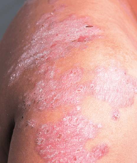
Metabolic syndrome affected skin expresses a greater level of inflammatory cytokines — tumour necrosis factor-α (TNF-α), interleukin 6 (IL-6), monocyte chemotactic protein-1(MCP-1), angiotensin, and other adipocytokines produced by adipocytes or the fat cells, which are now themselves recognized as a part of the innate immune system that regulates inflammation. Dysregulated production or secretion of these adipokines owing to adipose tissue dysfunction can contribute to the pathogenesis of obesity-linked complications. At the same time, these cytokines also cause epidermal proliferation and rejuvenation, but if the increased level of inflammatory cytokine persists, as it is in MetS, then rejuvenation of the barrier is difficult, and an inflammatory cascade can lead to systemic immune activation(3,8, 15–17). So there is no doubt that even if there are no documented clinical signs of the sensitive skin, we can’t disregard the subjective symptoms of patients, especially MetS patients.
In situations with pathological levels of TNF-α, this cytokine prevents proper barrier formation by inhibiting the expression of filaggrin and loricrin, resulting in a weakening of the skin barrier. IL-6 delays skin permeability barrier repair, wound healing, with delayed angiogenesis and collagen deposition(10). This explains why even a minimal external and/or internal irritation, which does not cause anything under normal circumstances, increases the skin’s sensitivity.
Oxidative stress
Oxidative stress, a condition of relative imbalance between reactive oxygen species (ROS) and antioxidants, is believed to play a central role in the pathogenesis of MetS(3).
The skin barrier is mediated by the skin’s xenobiotic biotransformation system, ROS-scavenging system, and excretory system, all of which contribute to the body’s total antioxidant capacity. Skin plays a role in the metabolism and elimination of xenobiotics, endogenous bioactive substances, lipids, and cholesterol(3, 18).
Xenobiotics such as exogenous chemicals, drugs, environmental pollutants, cosmetics, and dietary components form a major source of ROS. ROS are believed to activate proliferative and cell survival signalling that can alter apoptotic pathways that may be involved in the pathogenesis of a number of skin disorders. Antioxidants attenuate the damaging effects of ROS and can impair and/or reverse many of the events that contribute to epidermal toxicity and disease. However, increased or prolonged free radical action can overwhelm ROS defence mechanisms, contributing to the development of cutaneous diseases and disorders(3, 19). It is well recognized that inflammation is one manifestation of oxidative stress, and the pathways that generate the mediators of inflammation, such as interleukins, are all induced by oxidative stress(20).
“The skin barrier is mediated by the skin’s xenobiotic biotransformation system, ROS-scavenging system, and excretory system, all of which contribute to the body’s total antioxidant capacity.”
The various components of MetS show seasonal variations in their symptoms and signs: both blood pressure and blood cholesterol levels are increased in winter. Being more sensitive to environmental temperature than other organs, the possibility that the skin has a role in the association between blood pressure fluctuations and ambient temperature cannot be ruled out. A majority of sensitive skin sufferers report unpleasant sensory responses to cold temperatures and humidity, that cause lower water content in the stratum corneum and which, of course, is more often observed during the winter. So seasonal variations in MetS cases become more pronounced for sensitive skin patients(3, 4,21,22).
Endocrine abnormalities
Adipocytes secrete several endocrine peptides, such as leptin and adiponectin. These peptides play an important role in acute and chronic inflammatory processes through the regulation of cytokine expression that modulates the balance of helper T-cell types 1 and 2. As well as altered glucose metabolism and vascular endothelial biology(23).
Leptin is a hormone secreted by adipocytes. Most obese patients have elevated circulating leptin levels in the setting of functional leptin resistance. Leptin receptors have been located on tissues, including keratinocytes, fibroblasts, endothelial cells, and adipose tissue. Leptin is a key factor in regulating a wide range of biological responses, including energy homeostasis, hematopoiesis, neuroendocrine function, and immune responses(23).
It has a role in epidermal barrier development. Many studies have examined the beneficial role of leptin in wound healing, in injured skin — it promotes fibroblast proliferation and collagen synthesis, endothelial cell growth, and angiogenesis. However, at higher levels, as in metabolic syndrome patients, it proves toxic to the vasculature, leading to capillary leakage and avascular zones(8). In clinical cases, erythema, persistent or transient, and telangiectasia are observed in most cases in sensitive skin patients(7). So again, in sensitive skin patients suffering from MetS, these clinical signs are more pronounced.
Adiponectin plays an important role in metabolic function, inflammation and multiple biological activities in various tissues. Sebaceous glands express both adiponectin receptors (AdipoR) 1 and AdipoR2, and this finding suggested a role for adiponectin in sebum production. It promoted the formation of differentiated sebaceous gland structures with an increase in the number of sebaceous glands. And as we know, sebocytes lipids are secreted to the skin surface, where they play a major role in the skin barrier(24). But MetS patients have reduced adiponectin levels. In addition, its levels are inversely proportional to the number of Mets criteria — as the number of MetS criteria increases, adiponectin levels decrease, so barrier dysfunction accrues and skin sensitivity increases(25).
One recent study has proved that adiponectin is one of several downregulated genes in sensitive skin and reduced its expression and that of its receptors. Accompanied by low activated protein kinase activity, adiponectin is implicated in the development of sensitive skin(13).
Conclusion
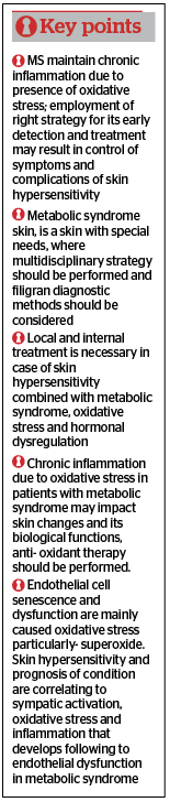
MetS affected sensitive skin has more pronounced skin barrier dysfunction, inflammation, immune response, oxidative stress, seasonal variations and weaker skin regeneration ability.
However, there is still a lack of information about the relationship between sensitive skin and metabolic disorders such as obesity, diabetes, and metabolic syndrome.
It remains unclear — whether metabolic syndrome causes sensitive skin or vice versa. Further research is needed.
Declaration of interest None
References
- The IDF work group. The IDF consensus worldwide definition of the metabolic syndrome. International Diabetes Federation, 2006.
- Wong ND. Metabolic syndrome: Cardiovascular risk assessment and management. Am J Cardiovasc Drugs. 2007;7:259–72
- Tanmay Padhi and Garima. Metabolic Syndrome and Skin: Psoriasis and Beyond. Indian J Dermatol. 2013 Jul-Aug; 58(4): 299–305.
- Farage, M.A., Maibach, H.I. Sensitive skin: closing in on a physiological cause. Contact Dermatitis, 2010; 62: 137–149.
- Misery, L., Jean-Decoster, C., Mery, S. et al. A New Ten-item Questionnaire For Assessing Sensitive Skin: The Sensitive Scale-10. Acta Derm Venereol, 2014; 94: 635–639
- Heinicke I.R., Adams D.H., Barnes T.M. and Greive K.A. Evaluation of a topical treatment for the relief of sensitive skin. Clin Cosmet Investig Dermatol. 2015; 8: 405–412
- Inamadar, A.C., Palit, A. Sensitive skin: An overview. Indian J Dermatol Venereol Leprol, 2013; 79: 9-16
- Yosipovitch G., DeVore A. and Dawn A. Obesity and the skin: Skin physiology and skin manifestations of obesity. J Am Acad Dermatol 2007; 56: 901-16
- Elias, P.M. Skin Barrier Function. Curr Allergy Asthma Rep. 2008 Jul; 8(4): 299–305
- Hänel, K.H., Cornelissen, C., Lüscher, B. et al. Cytokines and the Skin Barrier. Int. J. Mol. Sci, 2013; 14: 6720-6745
- Maibach, H.I., Tharp, M.D., Waldorf, H.A. The Essentials of Fundamental Skin Care: Scientific Rationale and Clinical Applications. Anatomic and physiologic aspects of the skin barrier
- Lowell, A., Goldsmith, Stephen, I., Katz, Barbara, A., Gilchrest et. al. Fitzpatrick`s dermatology in general medicine. Volume two. Eighth edition.. The McGraw-Hill. United States of America, 2012. 2576 lpp
- Fore-Pfliger, J. The epidermal skin barrier: implications for the wound care practitioner, part I. Advances in skin & wound care. 2004 Oct; 417-425
- Pons-Guiraud, A. Sensitive skin in dermatological practice. Dermfocus. Scientific review published by Avène Dermatological Laboratories, 2012; 46: 6-11
- Jason Lazar. Metabolic Syndrome: What It Is and Why You Should Treat It [online] – [posted: 04.10.2007]. Available: http://www.medscape.org/viewarticle/554276_print 3/
- Scarpellini E., Tack J. Obesity and metabolic syndrome: an inflammatory condition. Dig Dis. 2012; 30(2):148-53
- Zhou S.S., Li D., Zhou Y.M. and Cao J.M. The skin function: a factor of anti-metabolic syndrome. Diabetology & Metabolic Syndrome 2012, 4:15
- Janovska J., Kisis J., Voicehovska J. et al. Pilot study of early skin changes due to metabolic syndrome. March 2013.
- Mahjoub S and Masrour-Roudsari J. Role of oxidative stress in pathogenesis of metabolic syndrome. Caspian J Intern Med. 2012 Winter; 3(1): 386–396
- Farage, M.A., Katsarou, A., Maibach, H.I. Sensory, clinical and physiological factors in sensitive skin: a review. Contact Dermatitis, 2006; 55: 1–14.
- Farage, M.A., Mandl, C.P., Berardesca, E., Maibach, H.I. Sensitive Skin in China. Journal of Cosmetics, Dermatological Sciences and Applications, 2012; 2: 184-195
- Davidovici B.B., Sattar N., Jo¨rg P.C. et al. Psoriasis and Systemic Inflammatory Diseases: Potential Mechanistic Links between Skin Disease and Co-Morbid Conditions. Journal of Investigative Dermatology (2010) 130, 1785–1796
- Jung Y.R., Lee J.H., Sohn K.C. et al. Adiponectin Signaling Regulates Lipid Production in Human Sebocytes. PLoS One. 2017; 12(1): e0169824
- Von Frankenberg A.D., do Nascimento F.V., Gatelli L.E. et al. Major components of metabolic syndrome and adiponectin levels: a cross-sectional study. Diabetol Metab Syndr. 2014; 6: 26
- Kim EJ, Lee DH, Kim YK et al. Adiponectin Deficiency Contributes to Sensitivity in Human Skin. J Invest Dermatol. 2015 Sep; 135(9):2331-2334


