


Elastin
This protein is present in 2–4% of the dermis volume and dramatically changes between 30 and 70 years of age2.
Photoageing increases elastin storage owing to gene upregulation, which leads to a 5.3-fold increase in elastin expression7. This phenomenon is enhanced by reduced elastin degradation identified by the amount of racemised aspartic acid over time. From an immunohistochemical point of view, skin elastin and fibrillin are located in the papillary dermis, just below the basement membrane, and are small, oriented perpendicular to the epidermis. In the deep dermis, elastic fibres are thick and surround the vessels and annexa. With age, a rearrangement of these fibres is often observed2.
Fragmentation of elastic fibres induces a decrease in their number and diameter, and a biochemical modification of the fibres in polar amino acids, carbohydrates, lipids and calcium composition8.
Other proteins
Protein structure modifications are very common during ageing, with an increase in protein folding and a decrease in their aliphatic remnants exposed to water, especially in photoaged skin9. Amino acid composition of protein and free amino acids in aged skin differ significantly from that of young skin, and in older patients, an increase in their overall hydrophobicity is quite common.
Glycosaminoglycans
Glycosaminoglycans (GAGs) are specifically disaccharide units bound to a core protein (proteoglycans) or not (hyaluronic acid), and chondroitin sulfates, namely dermatan sulphate. They bind up to 1000 times their volume in water. With photoageing there is a paradoxical increase of GAGs, which stratify abnormally on the elastoic material of the superficial dermis, and not between collagen and elastic fibres as in normal skin. This phenomenon might explain the dry and leathery appearance of photoaged skin7.
Lipids
The stratum corneum is rich in corneocytes, which are embedded in a matrix of ceramides, cholesterol, fatty acids and smaller amounts of cholesterol sulphate, glucosylceramide and phospholipids in multilamellar sheets2. They create a waterproof barrier for the epidermis and quantitative variation of the ischaemic compounds induces xerosis and possible atopic dermatitis10. Many authors agree that the overall lipid content of human skin decreases with age10–12.
Skin detection and ageing
The techniques available for skin examination and its follow-up include histological analysis and more recently, direct immunohistochemical studies with regard to CD31 antigen and platelet endothelial cell adhesion molecule (PECAM) staining.
Immunohistochemistry is preferred to synthase phosphatase evaluation as it supplies semi-quantitative results. Intravital capillaroscopy can be performed either ex vivo or in vivo with luminescence microscopy and fluorescent angiography, photoplethysmography, and laser Doppler flowmetry for the arterial or dermal plexus.
Some dynamic tests such as finger immersion in cold water and finger circulation measurement show a blood-flow reduction with age progression, while in vivo fluorescine angiography showed an age-related decrease to the papillary dermis and little change in horizontal vessels.
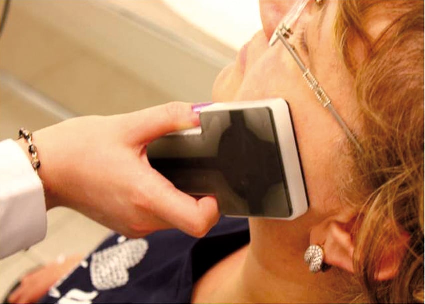
Figure 2 Skin Tester evaluation after filler injection
Epidermal thickness has been measured using confocal miscroscopy, or with ultrasound by Richard et al18, who performed an ultrasound analysis in 30 elderly women. Electrical conductance, colour, microrelief, skin thickness and subepidermal non-echogenic band (SENEB) were measured on the neck skin, which was damaged as a result of exposure to sunlight and on an adjacent part not exposed to the sun. Changes in SENEB, skin thickness, skin extensibility and elasticity, and colour heterogeneity were more evident in the sun-exposed skin, demonstrating that the cumulative effect of sun exposure can be the cause of atrophy or solar elastosis in older people.
Pellacani and Seidenari19 enrolled 40 women (aged 25–90 years) in order to study 12 different facial skin sites with a 20 mHz B echographic scanner, demonstrating an increase in facial skin thickness in old patients compared with the younger subjects.
To demonstrate a significant variation of skin parameters between different age groups, Seidenari et al20 performed an echographic evaluation of skin modifications on 48 patients (24 aged 27–30 years, and 24 over 60 years) with a 20 mHz B scanner at six different sites, showing an important regional variation of ultrasound reflection in older skin compared with the young skin. A consistent shift from low-intensity ultrasound echoes in the dermis of young subjects, to intermediate or high reflection amplitudes in older skin, was observed.
Gniadecka et al21 enrolled 10 older individuals (five men and five women; age range 74–87 years) and 10 control individuals (five men and five women; age range 22–29 years) to identify the role of structural protein ageing modifications to induce wrinkles, loss of elasticity and dryness. There was evidence that protein-specific amide I, amide III, and carbon–hydrogen stretching bands were shifted in photoaged forearm skin, suggesting an increase in protein folding. In contrast, significant changes were seen only in the amide I peak in chronologically aged skin. The intensity of the stretching band was increased in photoaged skin, but not in chronologically aged skin.
The same authors21 alsoinvestigated the water content and found that in young skin and chronologically aged skin, water was detectable in the bound form. In the photoaged skin, however, there was an increase in intensity, which reflects an increase in the non-protein-bound water (tetrahedron water clusters), concluding that proteins in photoaged skin are more compact and interact with water to a limited degree.

